

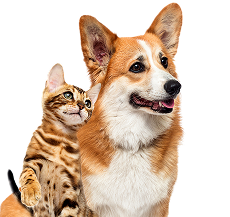

Animal Dermatology Services in Vancouver
Dogs
Breeds with >50% chance of food allergy vs environmental allergy: Request An...
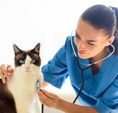
Cats
Cats experience a variety of skin and ear conditions that can cause itching, hair loss, infections, and discomfort.
Allergy Skin Testing
Allergies are a common and frustrating cause of skin issues in pets,...
READ MOREApoquel Treatment
Apoquel is a widely used medication designed to control itching and...
READ MOREFood Allergy Diagnosis
Food allergies in pets are a common but often misunderstood cause...
READ MOREAllergy Treatment
Allergies are among the most common causes of chronic skin problems...
READ MOREMites Treatment
Mite infestations are a common cause of itching, hair loss, and skin...
READ MOREEar & Nose Disorders
Ear and nose disorders are common problems in dogs and cats that can...
READ MOREHair Loss & Hormonal Disease
Hair loss, medically known as alopecia, is a common concern among pet...
READ MOREPaw & Nail Disorders
Paw and nail disorders are common yet often overlooked causes of...
READ MORESeborrheic & Breed Disorders
Seborrheic and breed-specific skin disorders are common causes of...
READ MOREPets Immune Disorder
Immune disorders affecting the skin are a complex group of diseases...
READ MORESkin Cancer & Tumors
Skin cancer and tumors are serious conditions that can affect dogs...
READ MORE